GASTROSCOPY

          Upper gastrointestinal endoscopy, also known as esophagogastroduodenoscopy (EGD, gastroscopy), is a minimally invasive procedure used to diagnose and treat diseases of the upper gastrointestinal tract, including the esophagus, stomach, and duodenum.

          This examination is performed using a flexible tube-shaped instrument called an endoscope, specifically a gastroscope. The device is equipped with a light source and a camera that transmits real-time images to a monitor. During the procedure, the gastroscope is gently advanced through the mouth and pharynx into the esophagus, stomach, and duodenum, allowing detailed visualization of the mucosa.

What is the purpose of gastroscopy?

EGD is performed to identify the cause of the following symptoms:

  • persistent heartburn;

  • gastrointestinal bleeding;

  • nausea and/or vomiting;

  • abdominal pain;

  • difficulty swallowing;

  • unexplained weight loss.

In addition, gastroscopy is used to diagnose a wide range of conditions, including:

  • gastroesophageal reflux disease (GERD);

  • fungal, eosinophilic, and other esophageal disorders;

  • gastritis (acute, chronic, atrophic, hyperplastic, autoimmune, and others);

  • peptic ulcer disease of the stomach or duodenum;

  • strictures or obstruction;

  • esophageal varices;

  • superficial inflammation, erosions, and ulcers;

  • benign and malignant neoplasms;

  • diaphragmatic hernias;

  • injuries caused by ingestion of caustic or toxic substances;

  • celiac disease;

  • precancerous conditions such as Barrett’s esophagus;

  • Crohn’s disease and other pathologies.

Therapeutic applications of gastroscopy

At the same time, gastroscopy can be used not only for diagnostic but also for therapeutic purposes. During the procedure, it is possible to perform:

  • endoscopic hemostasis for active bleeding;

  • removal of polyps and tumors;

  • dilation of narrowed segments;

  • extraction of foreign bodies;

  • placement of a gastrostomy tube;

  • ligation of esophageal varices;

  • sclerotherapy or glue occlusion of gastric or esophageal varices;

  • placement of metal or plastic stents;

  • placement of an intragastric balloon in patients with obesity.

EPK-i7010

Why Choose Gastro Health Center

1. Equipment

All procedures are performed using the state-of-the-art Japanese Pentax EPK-i7010 endoscopic system, providing 1080i HD+ imaging quality.
The OPTIVISTA Plus system with i-scan technology (SE, TE, OE) enhances image contrast and allows for detailed visualization of the mucosa.

2. Specialists

Our specialists undergo continuous training on international platforms and regularly participate in theoretical and hands-on workshops as part of continuous medical education programs.

3. Quality Indicators

  • Procedures are performed strictly according to clinical indications

  • Procedures are carried out exclusively under intravenous sedation

  • Complete photo documentation

  • Mandatory retroflexion examination

  • Use of simethicone

  • Examination time ≥ 7 minutes

  • Separate description of the Z-line and the gastroesophageal (GE) junction

  • Application of standardized classifications

  • Mandatory biopsy in cases of gastritis / H. pylori

  • Complication rate ≤ 0.2%

  • Endoscopist competency: ≥ 1000 diagnostic upper GI endoscopies (EGD)

  • Maintenance of competency: CME ≥ 20 hours per year

How to prepare for the examination?

Before undergoing gastroscopy, several important recommendations should be followed:

  • the examination should be performed according to a physician’s referral;

  • written informed consent may be required in certain cases;

  • the physician should be informed about any known allergies or sensitivities to medications or substances;

  • food and liquids should be avoided for at least 8 hours before the procedure, although additional dietary instructions may be provided when necessary;

  • it is important to inform the physician about possible pregnancy;

  • patients with bleeding disorders or those taking anticoagulants or other medications affecting blood clotting should notify the physician, as some drugs may need to be temporarily discontinued;

  • individuals with cardiovascular disease or immune system deficiency should inform the physician, since certain interventions may require prophylactic antibiotic therapy.

Gastroscopy may be performed under local anesthesia, using lidocaine spray in the oropharynx, or under intravenous sedation. The latter usually lasts 10–15 minutes and provides optimal comfort during the examination.


How to ensure a high-quality examination?

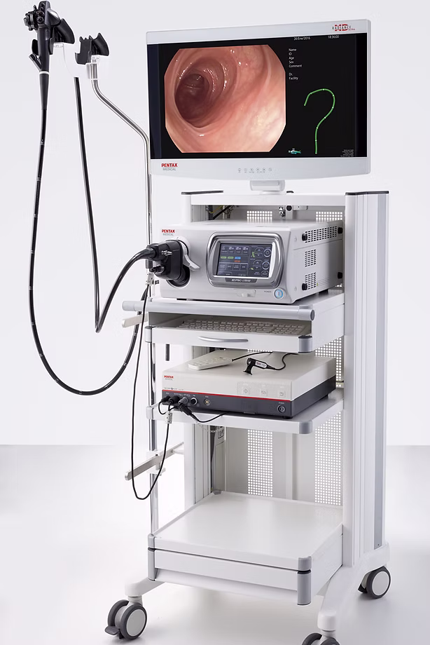
High-quality gastroscopy depends on proper patient preparation and strict adherence to medical recommendations. In addition, the level of equipment and the experience of the endoscopist play a crucial role.

Therefore, performing the examination under intravenous sedation allows for a more thorough inspection of the gastrointestinal mucosa and, if necessary, the safe performance of therapeutic interventions without causing discomfort to the patient.


What to expect after the examination?

After the procedure, the patient remains under medical observation for a short period. Once blood pressure, heart rate, and breathing are stable, the patient may leave the endoscopy unit.

However, if the examination was performed under intravenous sedation, driving a vehicle on the same day is strictly prohibited. Eating and drinking should be avoided until the gag reflex has fully returned. Mild throat discomfort or numbness may persist for several days and is considered normal.

If a biopsy specimen was obtained during the examination, histological results are typically available within 7–10 working days. Normal daily activities can usually be resumed unless otherwise advised by the physician.


Service prices

ONLINE BOOKING