ULTRASOUND EXAMINATION

Ultrasound examination (US) is a non-invasive imaging method used to visualize internal organs and tissues based on the reflection of ultrasound waves from anatomical structures. It is widely applied for primary diagnosis, dynamic monitoring, and assessment of treatment effectiveness.

Ultrasound is safe, does not involve ionizing radiation, and can be performed repeatedly, including in pregnant women and children.

Goals and diagnostic capabilities of ultrasound

Ultrasound allows evaluation of organ size and structure, detection of inflammatory and space-occupying lesions, fluid collections and calculi, assessment of blood flow (with duplex scanning), identification of functional disorders, and monitoring of disease progression.

Indications for ultrasound examination

Ultrasound is recommended in cases of pain of various localizations, digestive disorders, endocrine conditions, diseases of the genitourinary system, vascular pathology, suspicion of neoplasms, as well as for preventive examinations and follow-up assessment.

Types of ultrasound examinations performed at the clinic

We perform ultrasound examinations of the abdominal organs (including functional testing), pelvic organs, thyroid and parathyroid glands, breast, soft tissues, joints, salivary glands, and scrotum.

Folliculometry is also performed — dynamic ultrasound monitoring of the menstrual cycle for pregnancy planning.

Duplex vascular ultrasound

A combined ultrasound and Doppler examination used to assess vessel patency, blood flow direction, and hemodynamic characteristics. Applied for vessels of the upper and lower extremities, abdominal organs, and extracranial vessels.

Elastography (Sound Touch)

A modern ultrasound technique for quantitative assessment of tissue stiffness. It is used in suspected liver fibrosis, dynamic follow-up of chronic liver diseases, and evaluation of nodular lesions of the thyroid and breast. In certain clinical situations, it serves as a non-invasive alternative to biopsy.

Preparation for the examination

Preparation depends on the type of ultrasound study. For abdominal ultrasound, fasting for 6–8 hours is recommended. Requirements for other examinations are determined individually and clarified at the time of appointment scheduling.

Safety of the method

Ultrasound is a safe diagnostic modality, does not use ionizing radiation, and is suitable for repeated examinations.

When to see a doctor

In cases of pain of unclear origin, abnormal laboratory test results, chronic conditions requiring follow-up, or as part of preventive health screening.

ONLINE BOOKING
Ultrasound mindray resona i9

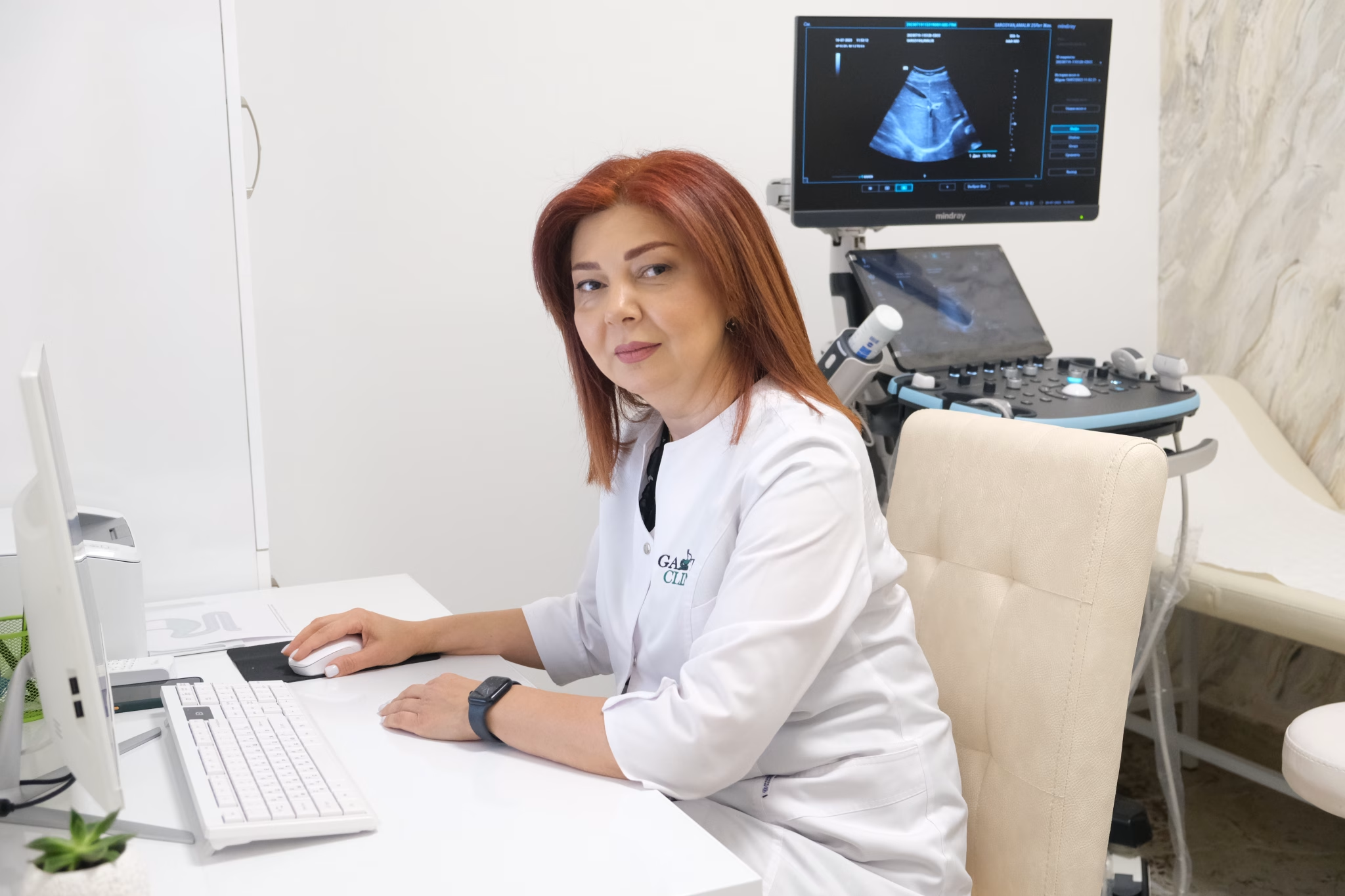
Why choose Gastro Health Center
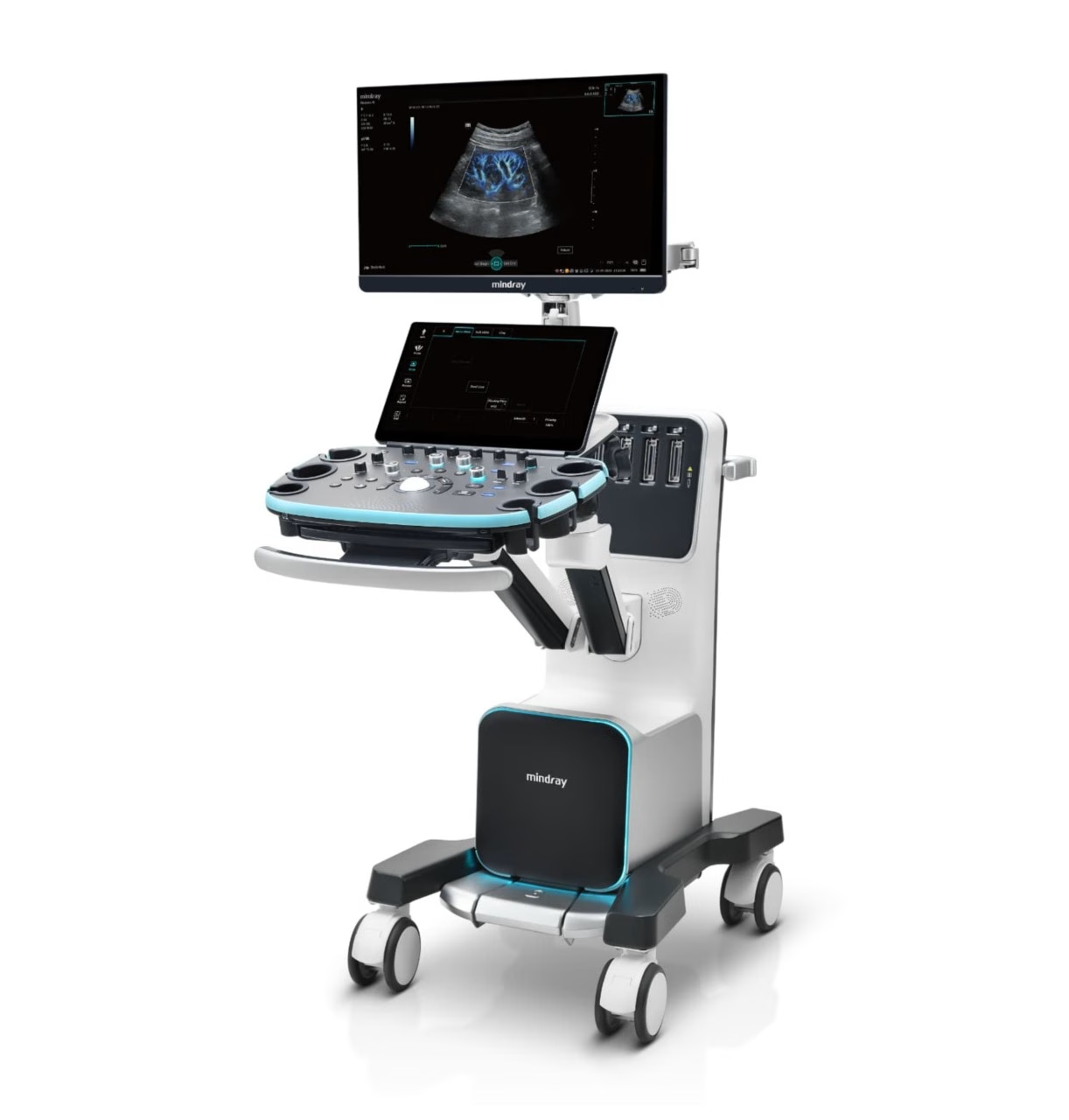
 

1. Equipment

Ultrasound examinations are performed using the expert-class Mindray Resona i9 system, equipped with the innovative ZST+ platform, providing high spatial and contrast resolution.

The system supports:

  • advanced Doppler modes, including V-Flow for vector blood flow assessment;

  • intelligent automated analysis tools:

    • Smart Breast (BI-RADS) for standardized breast evaluation;

    • Smart Thyroid (TI-RADS) for thyroid nodule assessment;

    • Smart HRI for quantitative hepatorenal index evaluation;

  • image optimization technologies (iClear, iBeam);

  • quantitative elastography (Sound Touch / HiFR STE).

2. Specialists

Ultrasound examinations are performed by physicians trained in diagnostic ultrasound with relevant clinical experience.

Our specialists regularly participate in continuing medical education (CME), are proficient in expert-level ultrasound systems, and interpret findings in accordance with current clinical guidelines and international classifications.

3. Quality indicators

  • Examinations are performed strictly based on clinical indications

  • Selection of technique, transducer, and scanning modes is individualized

  • Organs and structures are evaluated in multiple planes

  • Measurements follow international ultrasound standards

  • Mandatory image documentation of key anatomical structures and detected abnormalities

  • Use of standardized classifications and protocols (BI-RADS, TI-RADS, etc., when indicated)

For vascular studies, blood flow direction, velocity parameters, and spectral waveforms are recorded.
For elastography, quantitative and reproducible tissue stiffness measurements are obtained.

A structured ultrasound report is generated, reflecting the clinical question, findings, and diagnostic conclusion.

4. Safety and quality control

Ultrasound does not use ionizing radiation and is performed in accordance with the ALARA principle. Acoustic safety and technical condition of the equipment are monitored. Standardized protocols and physician expertise minimize incomplete or artifact-prone examinations.

5. What the patient receives

  • an informative and clinically meaningful examination;

  • an accurate report without excessive interpretation;

  • further management recommendations only when medically indicated.

Ultrasound team